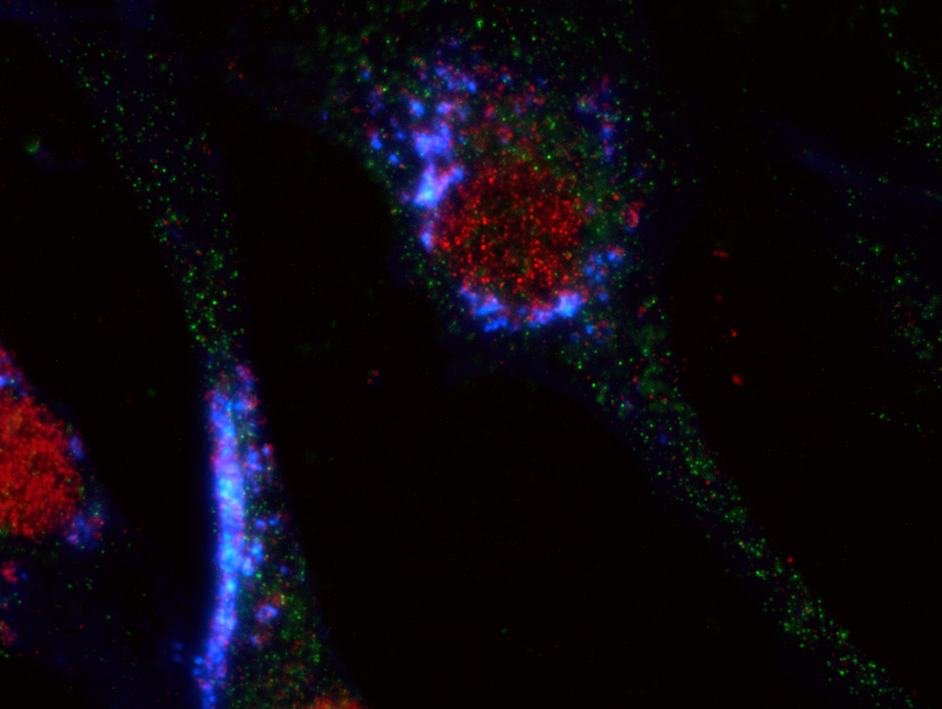
Because you were so consumed with longing to know what I do in lab all day, here’s a picture of some immunofluorescence staining I’ve been working on. Blue is cholesterol (labeled with filipin), which has accumulated in these cells as a result of treatment with a special drug. Red and green are two proteins I’m hypothesizing may experience changes as a result of the cholesterol accumulation (NPC1 and APP, respectively).
This is a single human fibroblast (skin) cell at 100x magnification. I imaged each channel separately and combined them in Photoshop with screen blending mode, adding individual levels adjustment layers to reduce background and make the signal more visible. This is helpful for visualizing where things are located within the cell, and if your fluorescent labels are colocalizing, that is, in approximately the same place. This could allow me to draw conclusions about which proteins are interacting with each other and where they move within the cell in response to stress (like cholesterol buildup).
A picture like this is the result of growing cells in culture for a number of days, permanently fixing them in place with chemicals, treating them with antibodies to label things I’m interested in with fluorescent tags, and a few hours under the microscope focusing and taking pictures. Filipin is especially difficult to image because the light required to excite it also rapidly burns it out. I get maybe a second of good blue glow before it’s gone for good and I have to find a new cell to image.
Immunofluorescence: Kind of like throwing stuff that glows in the dark into single cells and looking at them one hundred times larger than life. While the work itself gets tedious, seeing it through the lens never gets old. Go science!